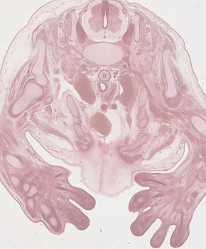
3D Fetal Atlas
Weeks gestation
All contents of the 3D Atlas of Human Embryology are licensed under the Attribution-NonCommercial-NoDerivatives 4.0 International (CC BY-NC-ND 4.0) license by Creative Commons.

This 3D embryo atlas contains a database of annotated 3D ultrasound images of early embryonal anatomy.
The atlas can only be viewed on a desktop or laptop. Browse the atlas by using the vertical menu on the left. Download the 3D-PDFs and use Adobe Reader X (or newer) on MS Windows or Mac OS for 3D interaction.
If you wish to use any of our images, please see our Citation Policy for information on how to do so. Images may only be used for non-commercial purposes.
We very much hope you will find this work useful. To help us improve our work, please share your feedback via the Contact Form.
If you would like to receive…. for …. purposes. Please make your request through the button below.
Histological sections (Amira or Tiff)
Amira files: Use Amira for combined use of the grey and label files. The label files also contain the label names.
3D-tiff files: IrfanView can be used for easy viewing of multipage tiff files. Use CTRL+PageDown/PageUp for scrolling trough the grey and label stack.
We are grateful to R.J. Cork and R.F. Gasser of the Virtual Human Embryo project (Available online at: http://virtualhumanembryo.lsuhsc.edu)
for kindly providing the photographs of the first series of sections of human embryos of the Carnegie collection, to E. Lockett and E. Wilson of the Human Developmental Anatomy Center, National Museum of Health and Medicine, US Army Garrison Forest Glen in Silver Spring MD for providing access to the Carnegie collection, and to G. Burton for generously providing the digital images of a stage 9 embryo of the Boyd Collection, scanned by A. Shelley.