The Human Organ Atlas Hub (HOAHub)
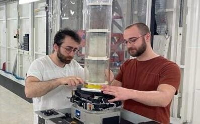
The Human Organ Atlas Hub (HOAHub) was launched September 21, 2023, with the first beamtime experiments beginning January 2024 at the European Synchrotron facility (ESRF).
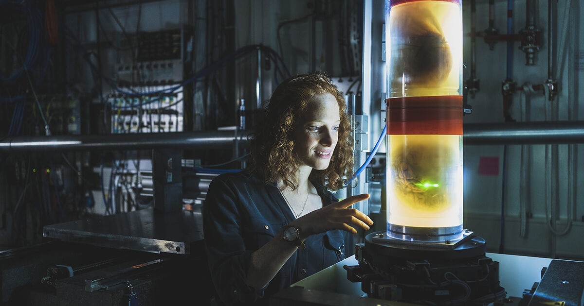
Professor Peter Lee and Dr Claire Walsh of UCL Mechanical Engineering, working as part of an international team from eight institutes including Dr. Bernadette de Bakker of Amsterdam UMC, have spearheaded an imaging research project aiming to use transformational X-ray tomography technology to scan whole human organs, and eventually our entire body, to an extremely high resolution – estimated resolution to be approaching one fiftieth of a human hair.
The group – comprised of a mixture of engineers, medics, educators, pathologists, cardiologists and more – have developed a new scanning technique collaboration with the ESRF, named Hierarchal Phase-Contract Tomography (HiP-CT). The technology enables whole intact organs to be examined at multiple scales all the way down to the cellular level. The scans can be created with a resolution of 25 microns, which is around twenty times the resolution of a clinical CT scanner and then small regions can be zoomed in on at <1 micron resolution, which is five hundred times the resolution of a CT scanner.
The team at UCL Mechanical Engineering partnered with Dr Paul Tafforeau from the ESRF, together with Drs Max Ackermann and Danny Jonigk from Aachen, to co-develop the technique. This international research project is funded by the European Union and the Chan Zuckerberg Initiative (CZI).

Background behind the HOAHub project
So far, the varying biomedical imaging techniques haven’t been able to show body parts in a detailed enough resolution. For example, computed tomography (CT) and magnetic resource imaging (MRI) both create images in millimetre resolution, whilst using a microscope to examine a biopsy only shows a tiny part of a whole organ. It has been impossible to create images of entire human organs with micro resolution – until now.
The solution to this problem initially stems from an imaging technique developed at the ESRF for palaeontology, with tomographic technology that scans fossils and ancient human skulls. This idea to use X-ray tomography technology was also assisted in 2020 by the development of the ESRF’s Extremely Brilliant Source (EBS). This created the brightest X-ray source ever (around 100 times brighter than before), which meant the larger soft samples like human organs could be scanned in a new level of detail.
Creating the Human Organ Atlas
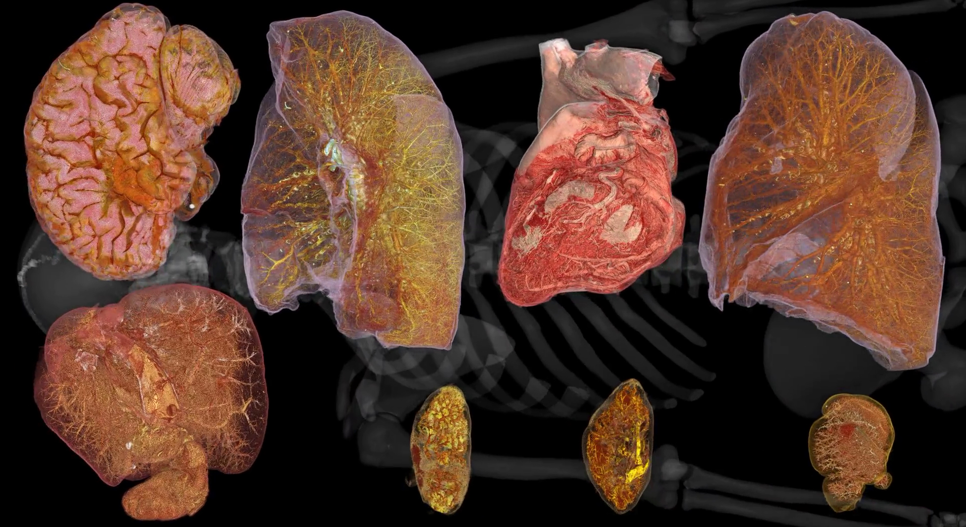
The project so far has created 3D images of over 50 organs, including lungs, brains, hearts, kidneys, spleens and livers. The aim is for this to become a reference database of organ images that anyone worldwide can access – explore images at human-organ-atlas.esrf.eu.
The HOAHub will enable an accelerated development of the HiP-CT technique and allow groups worldwide to use the technique. The HOAHub aims to solve key biomedical challenges and translate the findings to clinical applications and histology labs, as well as correlate up and down imaging scales and modalities. The HOAHub will create an open network of imaging by having all results open access for scientists, medics and students worldwide to use.
The vision behind the HOAHub was to create a synergistic and interdisciplinary group that exchanges ideas and resources (both physical and software-based) to create an efficient pipeline for Hierarchal Phase-Contract Tomography (HiP-CT). Ultimately, this project aims to answer some of the most important global biomedical questions.
Looking for Collaboration?
Are you a researcher or medical doctor with a compelling research question that aligns with the goals of the Human Organ Atlas HUB initiative? If so, we invite you to contact Dr. de Bakker or any of the Human Organ Atlas HUB Principal Investigators to discuss collaboration opportunities and apply for beam time. For more detailed information about the project and ways to collaborate, please visit the official Human Organ Atlas HUB website.
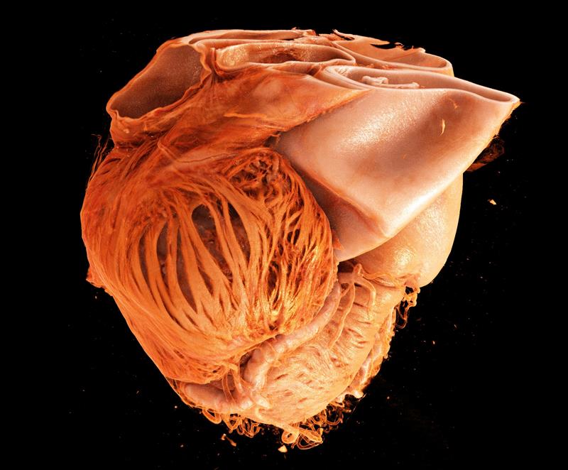
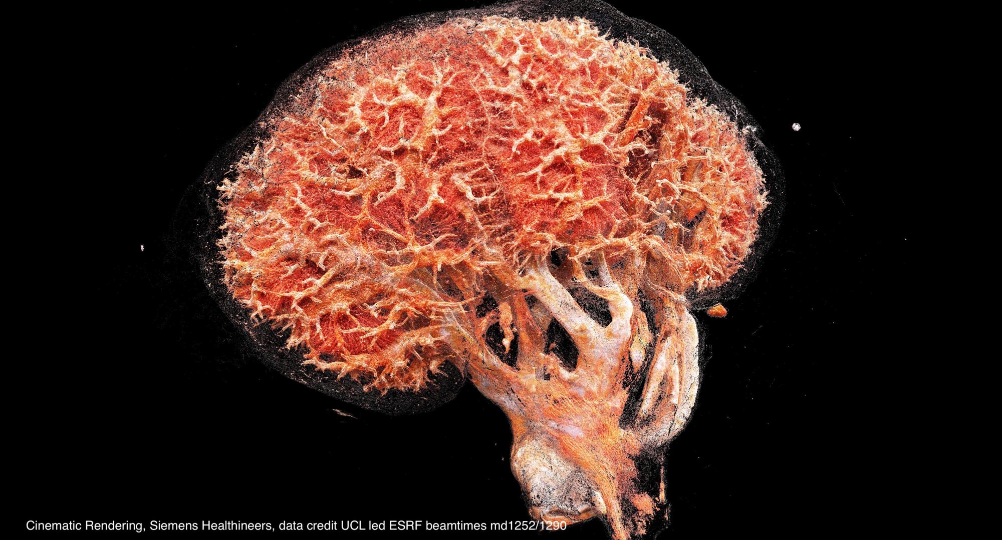
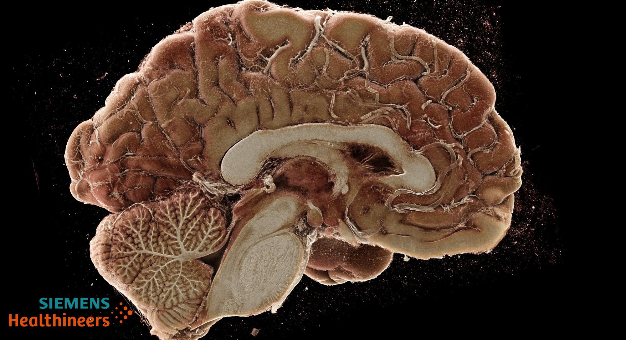
Images credit Klaus Engel, Cinematic Rendering, Siemens Healthineers, data credit UCL led ESRF beamtimes md1252/1290
Video produced by Paul Tafforeau. Data produced on the UCL led ESRF Beamtime MD1252, investigators PD Lee, CL Walsh, P Tafforeau et al. Click here for complete video.