The 4D Embryonic Brain Atlas
5-02-2026
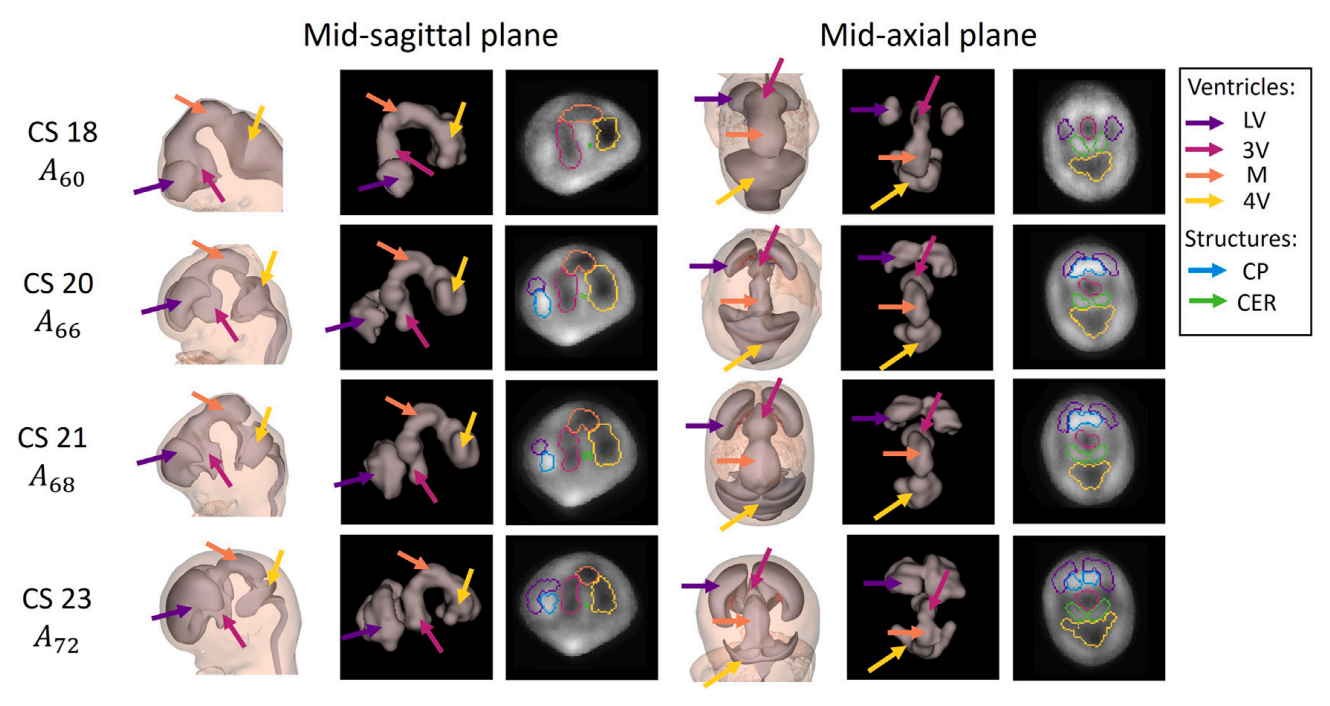
Dr. de Bakker and colleagues have published the 4D Human Embryonic Brain Atlas (8–12 weeks), a spatiotemporal atlas capturing rapid changes in brain anatomy during early development. The atlas was constructed from 831 high-quality 3D ultrasound scans across 402 pregnancies.
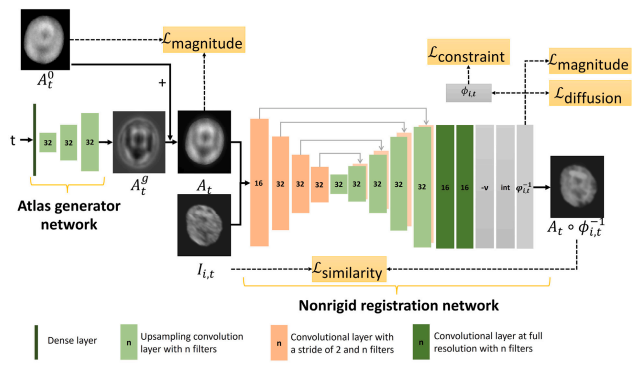
Published in Computerized Medical Imaging and Graphics (2026; Bastiaansen et al.), this work represents interdisciplinary collaboration at its best. AI specialists contributed expertise in registration, deep learning, and atlas generation, while the obstetrics and gynaecology team at Erasmus MC ensured consistent, high-quality first-trimester imaging.
The atlas holds particular importance for artificial intelligence in prenatal care. AI algorithms designed to detect and segment fetal organs are only as reliable as their ground truth data. Reference atlases such as this 4D brain atlas — alongside foundational resources like the 3D Embryo Atlas — provide essential training and validation datasets. Without robust ground truth, reliable clinical AI cannot be achieved.
Importantly, both the data and code are openly available, supporting transparency, benchmarking, and reproducibility in medical AI research.
This project exemplifies how developmental biology, clinical imaging, and artificial intelligence can converge to create tools with real clinical potential. By combining high-resolution imaging with computational innovation, the team moves closer to making AI in prenatal medicine not only technically advanced, but clinically meaningful.
Publication: Early thyroid development in Down syndrome
We are pleased to share a new publication in Human Molecular Genetics that sheds light on why thyroi…
Read moreA Milestone: 100 Scientific Publications!
I’m proud to share my 100th scientific publication: a review paper on an incredibly important yet st…
Read moreAdvancing the Human Organ Atlas at ESRF
I had the pleasure of joining the IAB meeting of the HOAHub at the ESRF – The European Synchrotron i…
Read more