
3D Synchrotron X-Ray Imaging of Uterine Vasculature in Adenomyosis
3-11-2025
We are proud to share our latest publication: “Revealing the Unseen: 3D Synchrotron X-Ray Imaging of Uterine Vasculature in Adenomyosis”, published in Angiogenesis.
Adenomyosis is a uterine disorder that affects one in five women. Symptoms such as menstrual disorders and reduced fertility can have a profound impact on quality of life, yet the underlying causes of adenomyosis remain unknown. Histologically, the condition is characterized by the invasion of the inner uterine layer into the outer layer, inducing the formation of new blood vessels (angiogenesis).
In this paper, we present a proof-of-principle for an innovative 3D imaging technique: synchrotron-based Hierarchical Phase-Contrast Tomography (HiP-CT). For the first time, this approach allowed us to visualize and reconstruct the complete microvasculature of a uterus affected by adenomyosis without the need for physical sectioning. The resulting high-resolution 3D reconstructions provide a powerful new tool for studying uterine disorders.
These initial findings represent just a glimpse of the potential of HiP-CT data. In future work, we aim to use this technique to quantify vascular differences between healthy and adenomyotic uteri, with the goal of advancing our understanding of the disorder’s etiology.
As members of the Human Organ Atlas Hub (HOAhub), we have unique access to the European Synchrotron Radiation Facility (ESRF). Its exceptional phase-contrast imaging capabilities enable highly detailed visualization of soft biological tissues, opening new frontiers in organ-level research.
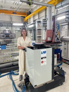
This work was led by Veerle Michels (see picture), first author of the paper. After completing her studies in Technical Medicine, she started as a PhD candidate under the supervision of Prof. Judith Huirne and Dr. Bernadette de Bakker. Achieving a publication of this quality in Angiogenesis during the very first year of her PhD is an impressive accomplishment and highlights the strength and potential of her research trajectory.
We thank all members of the HOAhub uterine innervation team for their invaluable contributions to this work.
👉 Read the full publication in Angiogenesis
Publication: Early thyroid development in Down syndrome
We are pleased to share a new publication in Human Molecular Genetics that sheds light on why thyroi…
Read moreA Milestone: 100 Scientific Publications!
I’m proud to share my 100th scientific publication: a review paper on an incredibly important yet st…
Read moreThe 4D Embryonic Brain Atlas
Dr. de Bakker and colleagues have published the 4D Human Embryonic Brain Atlas (8–12 weeks), a spati…
Read more