Chapter Publication: Advanced Imaging in Fetal and Embryonic Research
6-05-2025
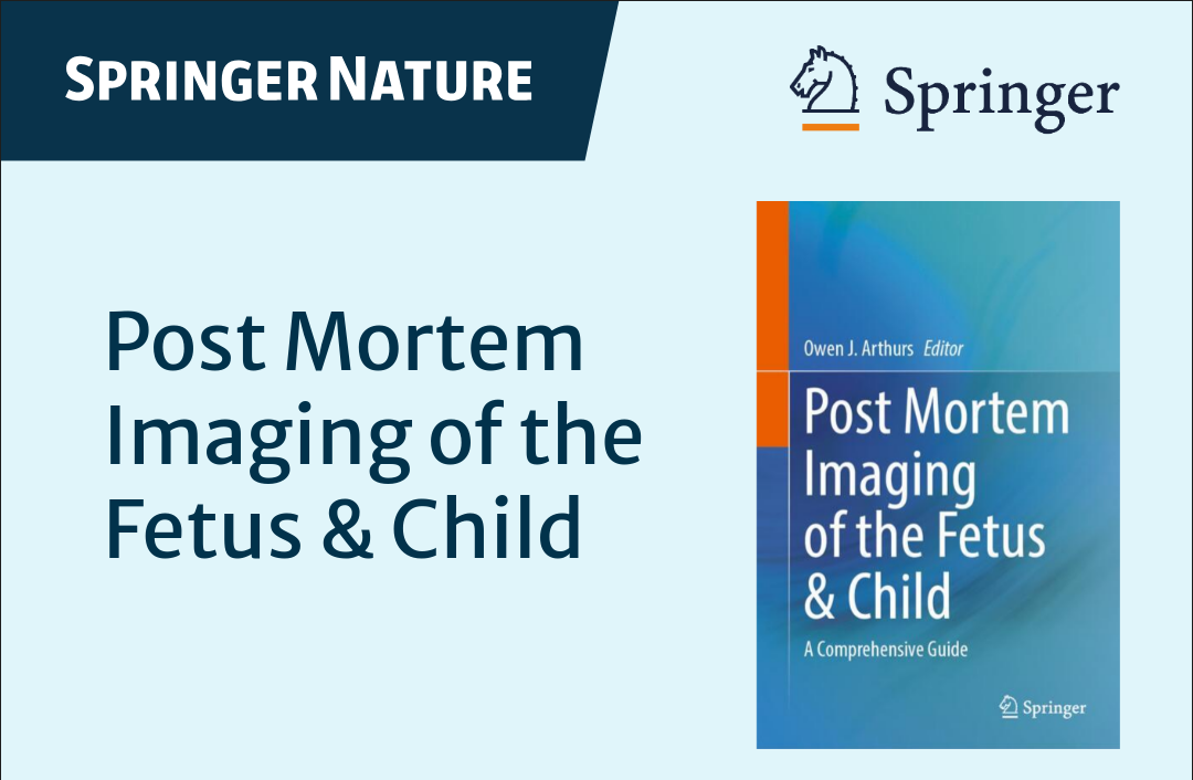
The book Post Mortem Imaging of the Fetus & Child, edited by Owen Arthurs, Professor of Radiology, Great Ormond Street Hospital for Children (London), has just been published!
Yousif Dawood and Bernadette de Bakker co-authored Chapter 6: Advanced Imaging Techniques, together with Gustav Strijkers, Ian Simcock and Michael Aertsen. In this chapter, they dive into cutting-edge, non-invasive imaging modalities—micro-CT and ultra-high field MRI—for studying human embryos and fetuses post mortem. They explore their technical principles, applications, and protocols, and compare their strengths in visualizing delicate fetal structures at high resolution. These techniques offer promising alternatives to traditional autopsy, especially for early gestational ages. They also touch on exciting future directions, including artificial intelligence and synchrotron imaging.
This book serves as a reference standard for anyone wanting to begin or improve their post mortem imaging in children, including pathologists, radiologists, radiographers, technicians, mortuary staff, fetal medicine & obstetricians, forensic imaging staff and pathologists. Divided into three parts, the book covers imaging principles and techniques, interpretation of post mortem imaging, and guidance on which imaging technique to use for which clinical scenarios.
More info about the book can be found here.
Thanks to Owen Arthurs for the opportunity and excellent collaboration—and to the coauthors for their inspiring contributions throughout this project.
Publication: Early thyroid development in Down syndrome
We are pleased to share a new publication in Human Molecular Genetics that sheds light on why thyroi…
Read moreA Milestone: 100 Scientific Publications!
I’m proud to share my 100th scientific publication: a review paper on an incredibly important yet st…
Read moreThe 4D Embryonic Brain Atlas
Dr. de Bakker and colleagues have published the 4D Human Embryonic Brain Atlas (8–12 weeks), a spati…
Read more