3D Embryo Neurovascular Atlas Published!
23-08-2026
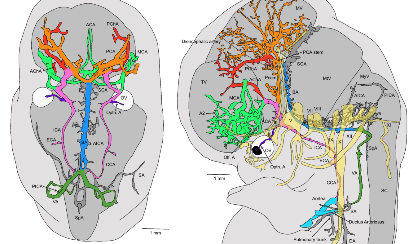
Karl Jacobs and colleagues have published a new paper in Life: Vascular Neuroembryology: The Genesis of the Intracranial Arterial System and the Circle of Willis. Alongside the paper, the team also launched the 3D Embryo Neurovascular Atlas, a new open resource that makes these reconstructions available to the wider research community.
Using stage-resolved 3D reconstructions of the developing intracranial arteries, based on serially sectioned human embryos from the 3D Embryo Atlas, the study traces how the circle of Willis gradually takes shape during the earliest weeks of development. By comparing these reconstructions to the classical embryological literature, the team refines several long-standing assumptions about the timing and pattern of vascular development in the head, particularly around the eye and the trigeminal region. Understanding this developmental process may help explain why so much anatomical variation in the adult circle of Willis exists, variation that is clinically relevant in conditions such as stroke and aneurysms.
Beyond its scientific value, the atlas also has a strong educational purpose. Anatomy teachers, students, and clinicians can explore the reconstructions as interactive 3D-PDFs, which can simply be downloaded and viewed, making it easy to study the developing vasculature layer by layer rather than through static images alone.
The work was carried out together with Dr. Bernadette de Bakker and Prof. Roelof-Jan Oostra, along with colleagues from ACTA, Amsterdam UMC, Erasmus MC, and other collaborating institutes. This research was made possible in part by the AR&D grant awarded to Karl earlier this year, as well as generous funding from the De Snoo–van ‘t Hoogerhuijs Foundation.
Congratulations to Karl and the entire team on this milestone!
Questionnaire: Help us understand pregnancy loss
Every year, millions of parents around the world experience the loss of a pregnancy, and yet, within…
Read moreESHRE 2026 in London
This July, we attended ESHRE 2026 in London, one of the world’s leading events in reproductive scien…
Read moreISFRI & IAFS 2026 in Sofia, Bulgaria
This year’s International Society of Forensic Radiology and Imaging (ISFRI) meeting joined forces wi…
Read more