Review Imaging Fetal Anatomy
27-11-2022
Super proud at my team and fellow researchers for publishing our review entitled ‘Imaging Fetal Anatomy‘ in Seminars in Cell & Developmental Biology!
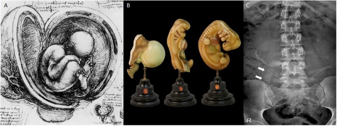
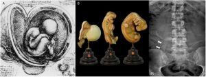
“The aim of this review is to provide an overview of the past, present and future techniques used to capture images of the developing human embryo and fetus and provide the reader newest insights in upcoming and promising imaging techniques. The reader is taken from the earliest drawings of da Vinci, along the advancements in the fields of in utero ultrasound and MR imaging techniques towards high-resolution ex utero imaging using Micro-CT and ultra-high field MRI. Finally, a future perspective is given about the use of artificial intelligence in ultrasound and new potential imaging techniques such as synchrotron radiation-based CT to increase our knowledge regarding human development.”
Thanks Yousif Dawood, Marieke Buijtendijk, Harsha Shah, Hans Smit, Karl Jacobs, Jaco Hagoort, Roelof-Jan Oostra, Professor Tom Bourne and Maurice van den Hoff!
“Clear images of embryonic and fetal development can also be used in training for sonographers and fetal surgeons, or to educate parents expecting a child with a fetal anomaly.”
Questionnaire: Help us understand pregnancy loss
Every year, millions of parents around the world experience the loss of a pregnancy, and yet, within…
Read more3D Embryo Neurovascular Atlas Published!
Karl Jacobs and colleagues have published a new paper in Life: Vascular Neuroembryology: The Genesis…
Read moreESHRE 2026 in London
This July, we attended ESHRE 2026 in London, one of the world’s leading events in reproductive scien…
Read more