3D Ultrasound Atlas
Comparative fetal anatomy
Below are annotated images of a fetus at 11 weeks gestation, imaged in-vivo using 3D ultrasound imaging with CrystalVue™ and RealisticVue™ applied.
This specimen was matched to a Carnegie Stage 23 (CS23) embryo from the Carnegie Collection. Histological sections and a 3D embryological model of this CS 23 specimen has been used and adapted from B.S. de Bakker (Science), with permission and are used as a reference standard to validate and annotate anatomical the structures visualised using 3D ultrasound imaging.
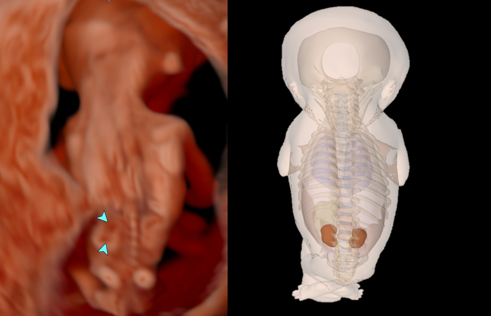
Coronal view of fetus demonstrating the fetal kidneys, comparable to gestation-matched (Carnegie Stage 23) embryological model in the same plane
Image credit for the right image: 3D Embryo Atlas, B.S. de Bakker et al., An interactive three-dimensional digital atlas and quantitative database of human development, Science 2016; 354
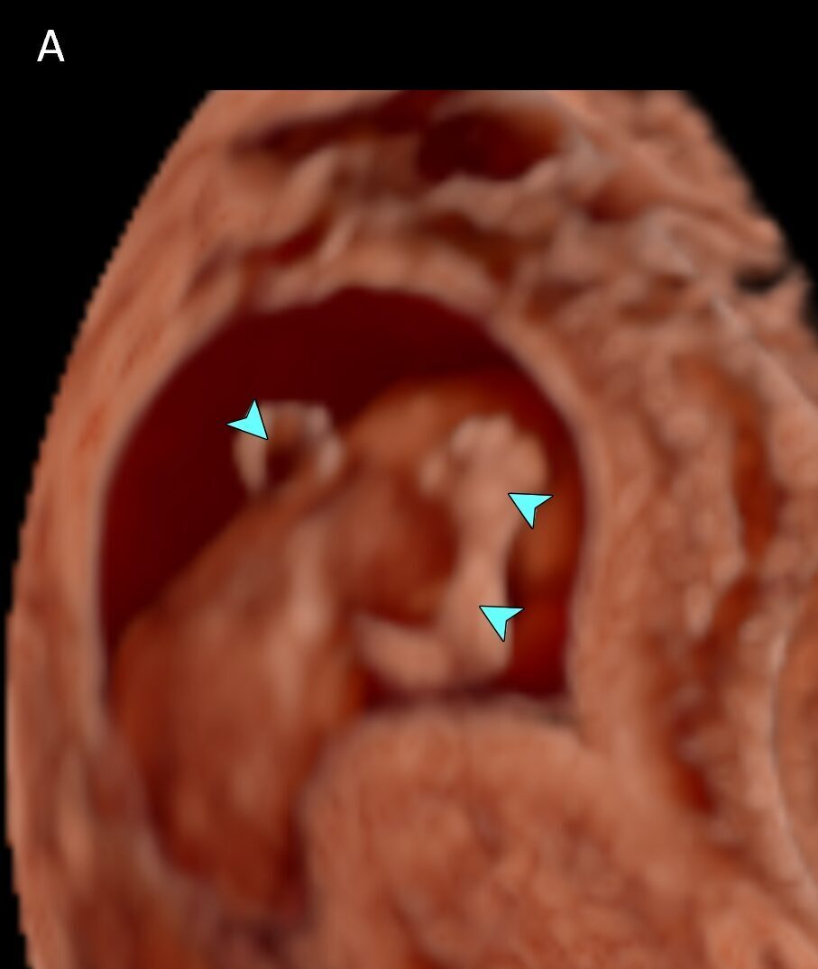
Arm – fingers
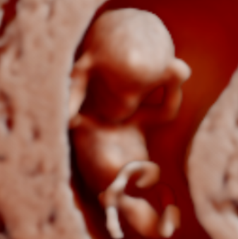
Face – Nasal tip
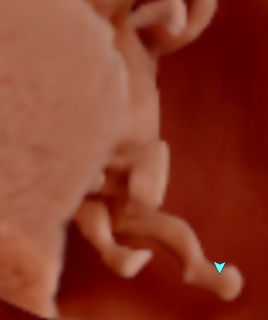
Legs – feet
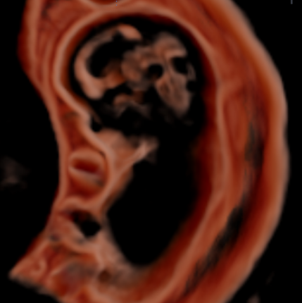
Ventricles in sagittal section
The images above show the 3D rendered volume with CrystalVue™ and RealisticVue™ applied.