Comparative fetal anatomy
Below are annotated images of a fetus at 8 weeks gestation, imaged ex-vivo using 3D ultrasound imaging with CrystalVue™ and RealisticVue™ applied (CRL: 2.50cm; estimated gestational age 8+6 based on morphological characteristics; range 8+6 – 9+6 weeks).
This specimen was matched to a Carnegie Stage 21 (CS21) embryo from the Carnegie Collection (CRL: 1.73cm; estimated gestational age 8+2, range 7+4 – 8+6 weeks based on CRL). Histological sections and the 3D-models have been adapted with permission from de Bakker et al., 2016 and were used as a reference to validate and annotate the anatomical structures visualised using 3D-ultrasound imaging.
The arrows in each image indicate anatomical structures. You can view the interactive structure annotations by moving your mouse over the arrows.
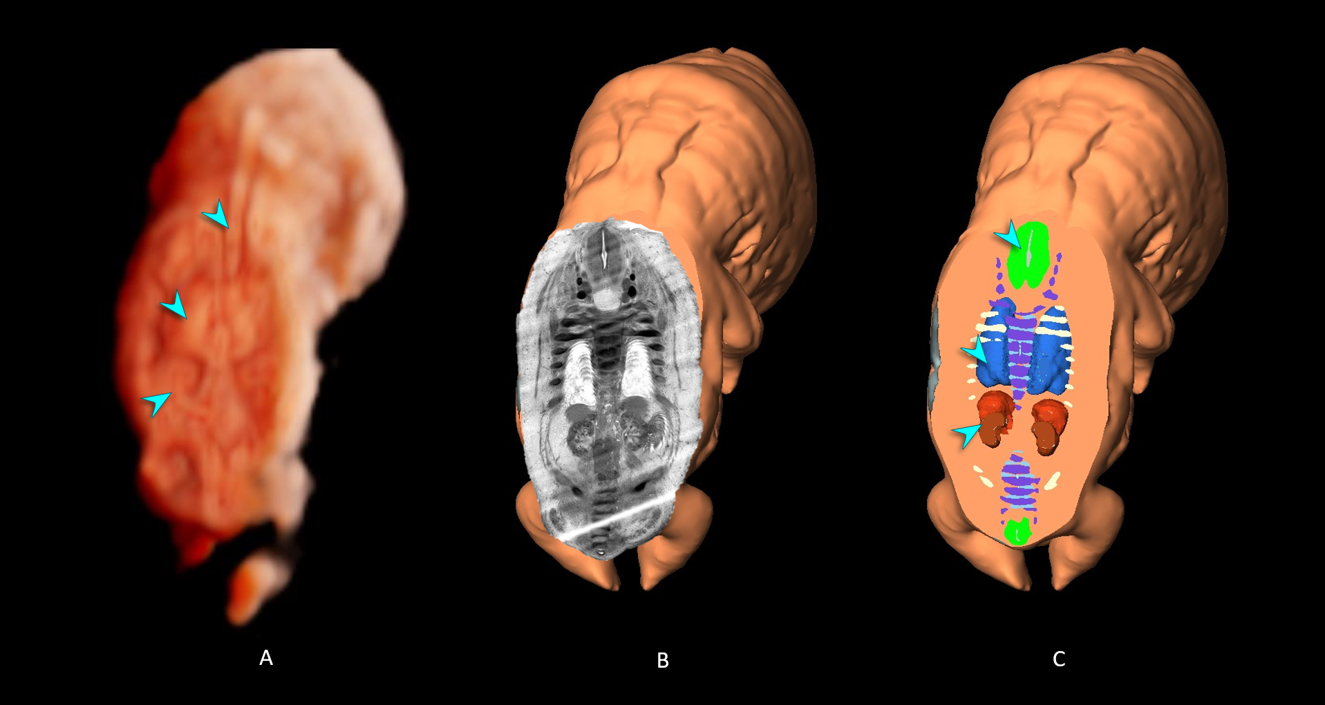
- 3D ultrasound volume with CrystalVue™ and RealisticVue™ rendering. The volume was sectioned in a coronal plane to visualise internal structures.
- Histological section from the Carnegie Collection. A matching imaging plane was selected to validate the structures visualised. The original specimen was sectioned in the transverse plane. The sections were imported into a digital post-processing software (Amira, ThermoFisher Scientific) and aligned. From this imaging stack, the software can recreate other imaging planes, including the coronal imaging plane as demonstrated here.
- Cross section of a 3D embryological model based on histological sections as shown in image B, the cross section was taken in the same imaging plane as image B.
*The exact border between the adrenal glands and kidneys cannot be clearly distinguished due to the small size of the structures and limits of ultrasound resolution.
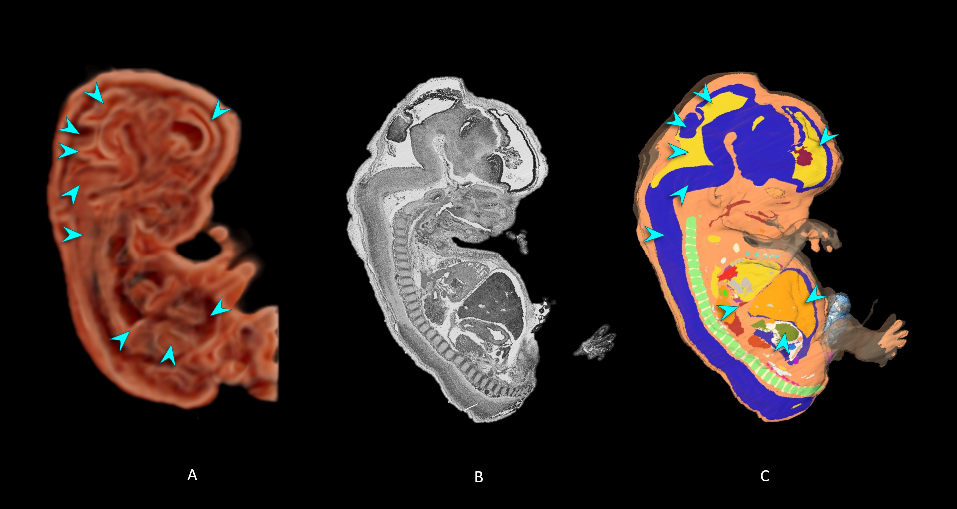
- 3D ultrasound volume with CrystalVue™ and RealisticVue™ rendering. The volume was sectioned in a parasagittal plane to visualise internal structures.
- Histological section of the matched specimen from the Carnegie Collection (reconstructed imaging plane).
- Cross section of a 3D embryological model based on histological sections as shown in image B, the cross section was taken in the same imaging plane as image B.
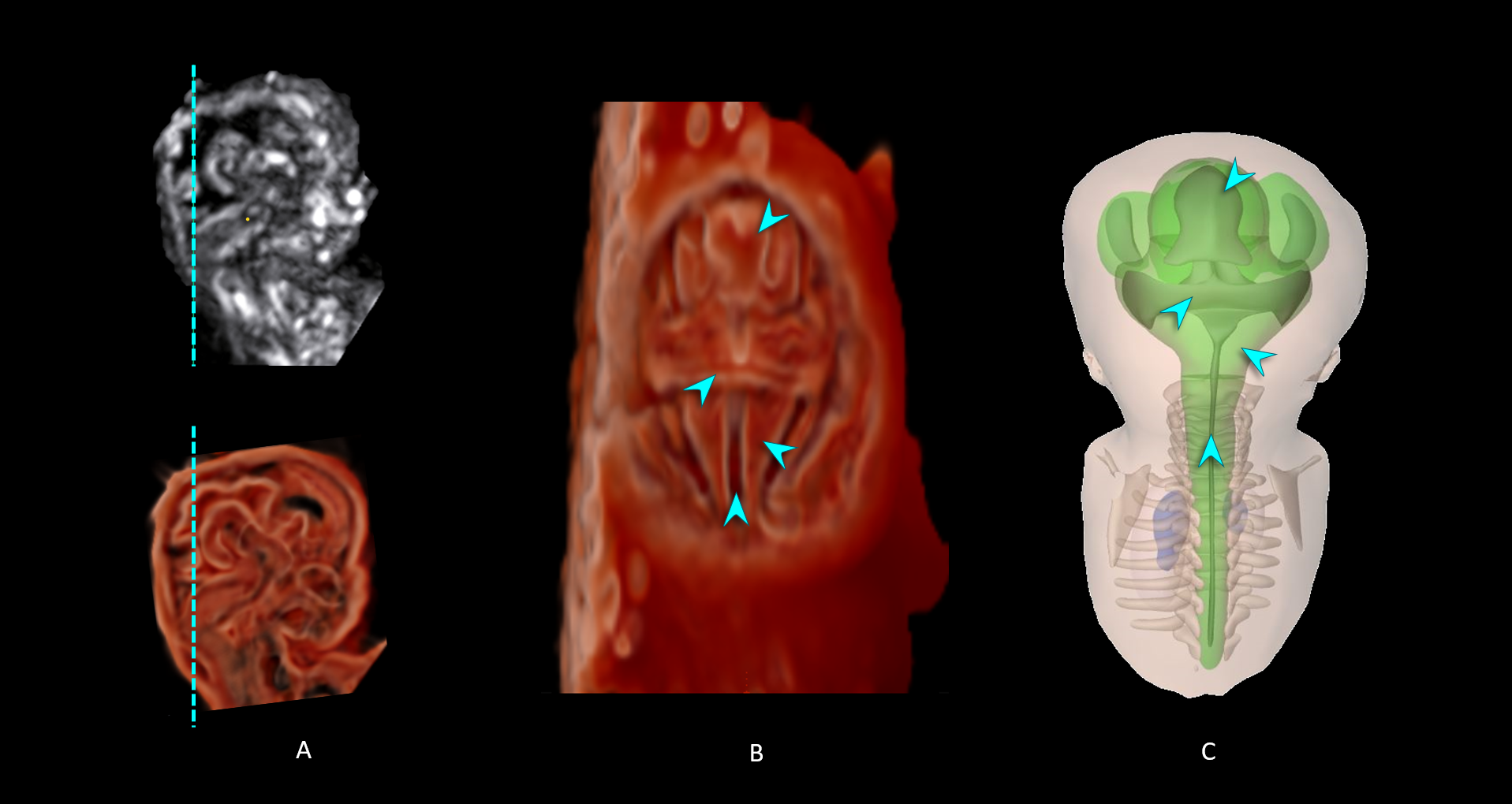
- 3D ultrasound volume of the head and brain. The top image shows the reconstructed 2D sagittal plane selected from the volume in multiplanar mode. The bottom image shows the 3D rendered volume with CrystalVue™ and RealisticVue™ applied, which was inverted to better visualise intracranial structures.
- The same 3D ultrasound volume presented in panel A was turned 45 degrees to view the back of the head. The volume was sectioned in a coronal plane to visualise structures located in the posterior cranial fossa. The dotted lines in panel A indicate the sectioning plane.
- 3D embryological model showing the same structures as those visualised in panel A.