Comparative fetal anatomy
Below are annotated images of a fetus at 9 weeks gestation, imaged ex-vivo using 3D ultrasound imaging with CrystalVue™ and RealisticVue™ applied (CRL: 3.14cm; estimated gestational age 9+6 weeks, range 9+2 – 10+4 weeks).
This specimen was matched to a Carnegie Stage 23 (CS23) embryo from the Carnegie Collection (CRL: 3.0cm; estimated gestational age 9+6, range 9+1 – 10+4 weeks). Histological sections and the 3D-models have been adapted with permission from de Bakker et al., 2016 and were used as a reference to validate and annotate the anatomical structures visualised using 3D-ultrasound imaging.
The arrows in each image indicate anatomical structures. You can view the interactive structure annotations by moving your mouse over the arrows.
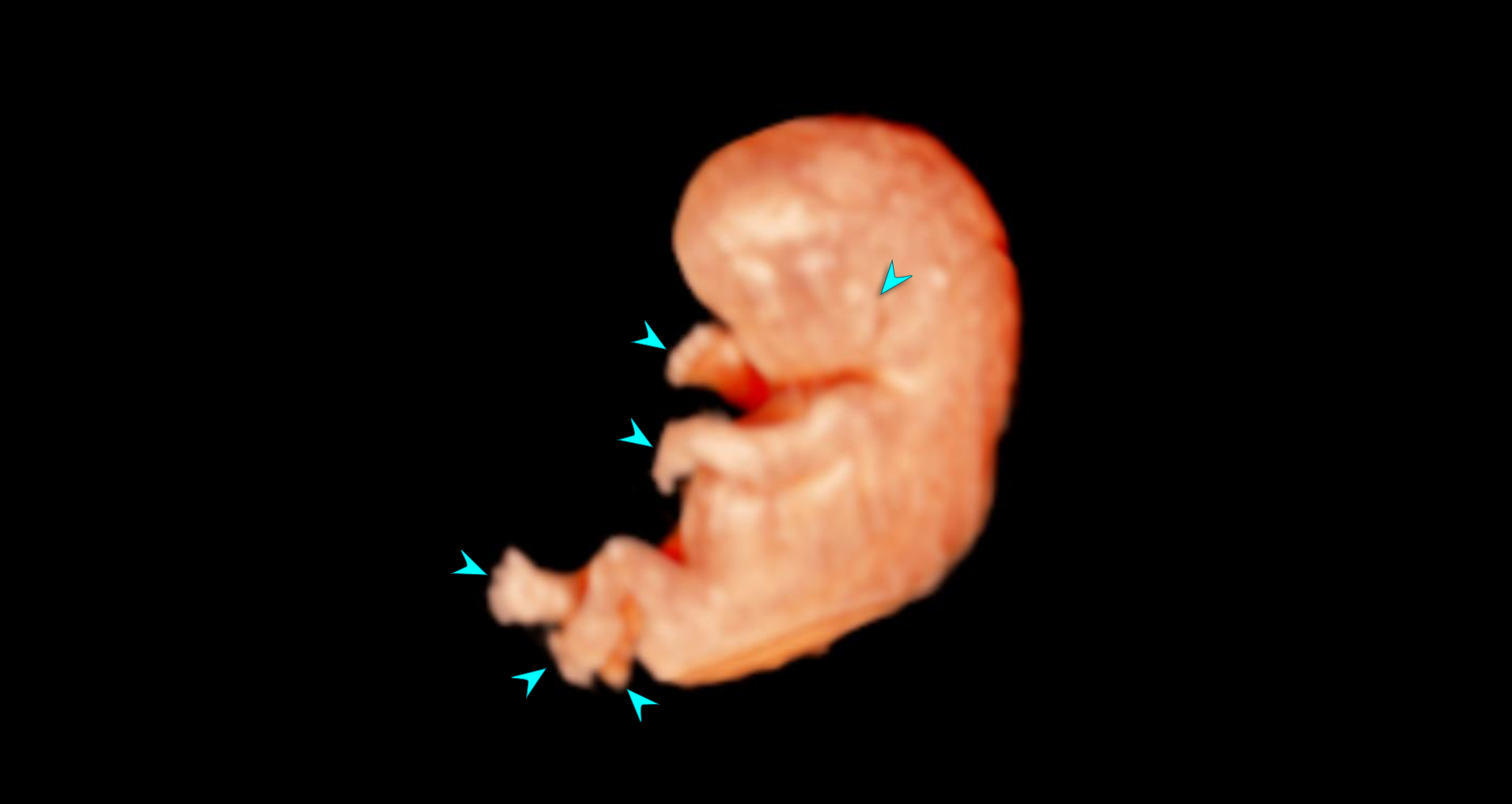
Surface view of a 3D ultrasound volume with CrystalVue™ and RealisticVue™ rendering applied.
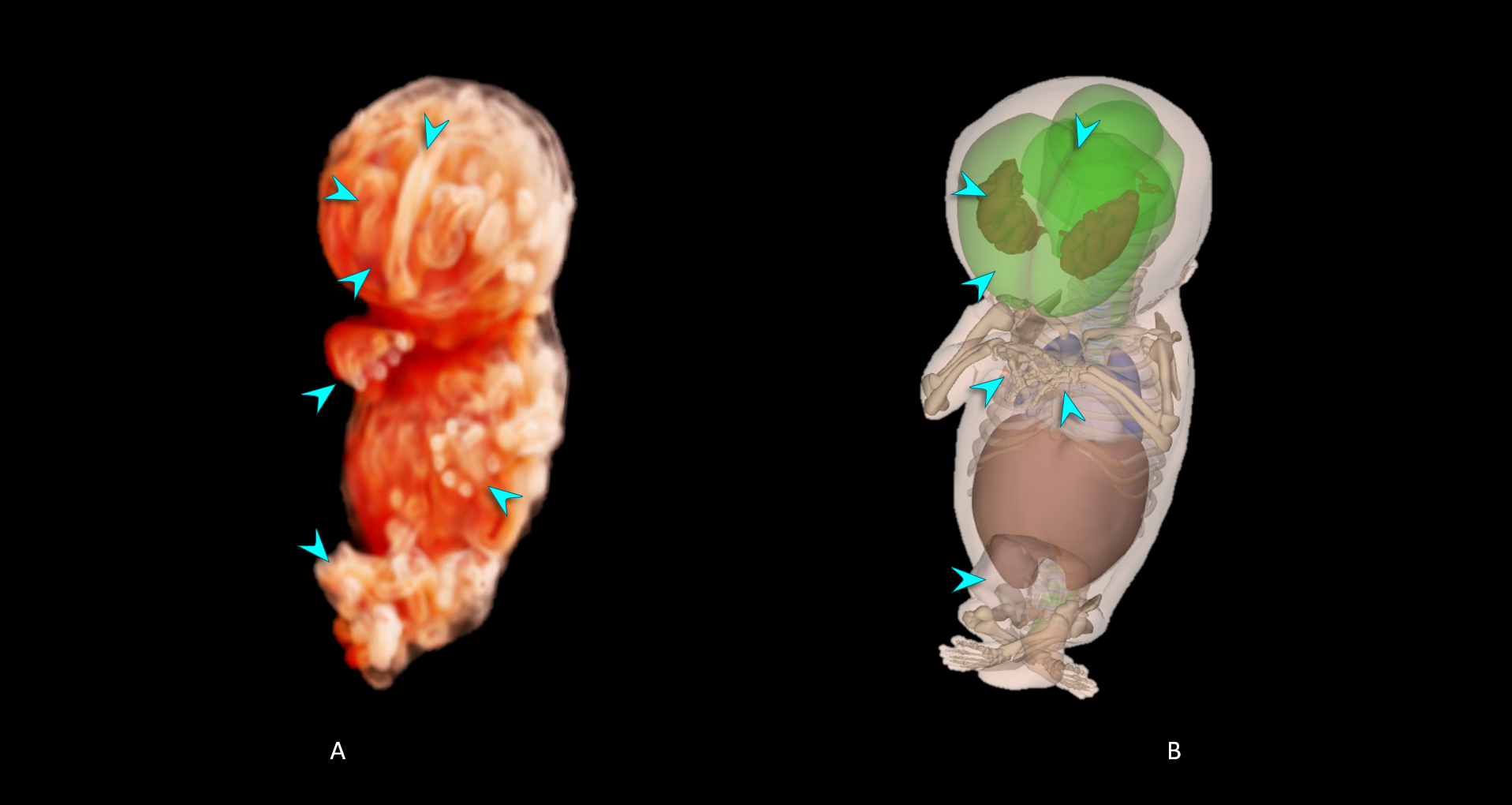
- 3D ultrasound volume with CrystalVue™ and RealisticVue™ rendering. The level of CrystalVue™ complexity and transparency have been increased and the choroid plexus and lateral ventricles are visualised.
- 3D embryological model showing the same structures as those visualised in A.
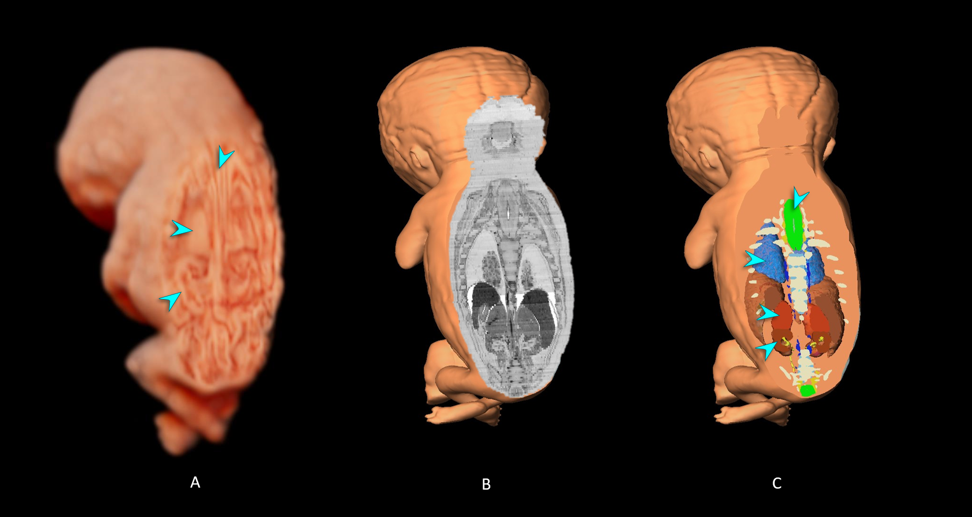
- 3D ultrasound volume with CrystalVue™ and RealisticVue™ rendering. The volume was sectioned in a coronal plane to visualise internal structures.
- Histological section of the matched specimen from the Carnegie Collection. A matching imaging plane was selected to validate the structures visualised. The original specimen was sectioned in the transverse plane. The sections were imported in to a digital post-processing software (Amira, ThermoFisher Scientific) and aligned. From this imaging stack, the software can recreate other imaging planes, including the coronal imaging plane as shown in this image.
- Cross section of a 3D embryo model based on histological sections as shown in image B, the cross section was taken in the same imaging plane as image B.
*The exact border between the adrenal glands and kidneys is not clearly distinguishable due to their small size and limits of ultrasound resolution.
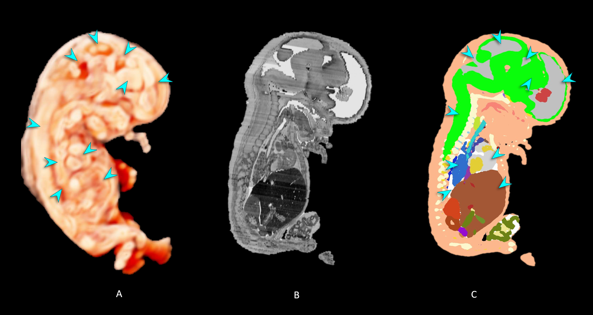
- 3D ultrasound volume with CrystalVue™ and RealisticVue™ rendering. The volume was sectioned in a parasagittal plane to visualise internal structures.
- Histological section of the matched specimen from the Carnegie Collection (reconstructed imaging plane).
- Cross section of a 3D embryo model based on histological sections as shown in image B, the cross section was taken in the same imaging plane as image B.