Comparative fetal anatomy
Case 1
Below are annotated images of a healthy fetus – 13 weeks and 5 days (13 + 5 weeks) gestation – imaged ex-vivo using 3D ultrasound with CrystalVue™ and RealisticVue™ rendering. Thereafter, the specimen was imaged using contrast-enhanced microCT which was used as an anatomical reference standard. The ultrasound and microCT images are directly compared, allowing for validation and annotation of the the structures visualised in 3D ultrasound imaging.
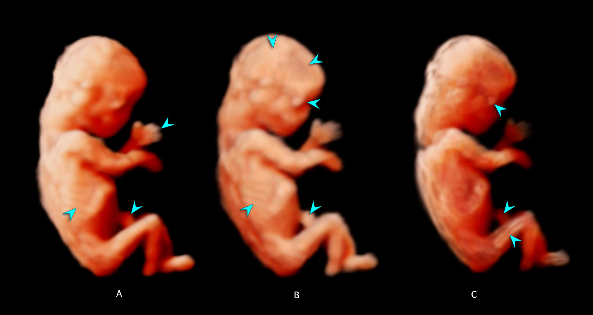
- – C. Increasing levels of CrystalVue™ complexity and transparency are applied to the 3D ultrasound volume thereby highlighting different (internal) structures.
- 3D ultrasound volume with CrystalVue™ and Realistic Vue™ applied.
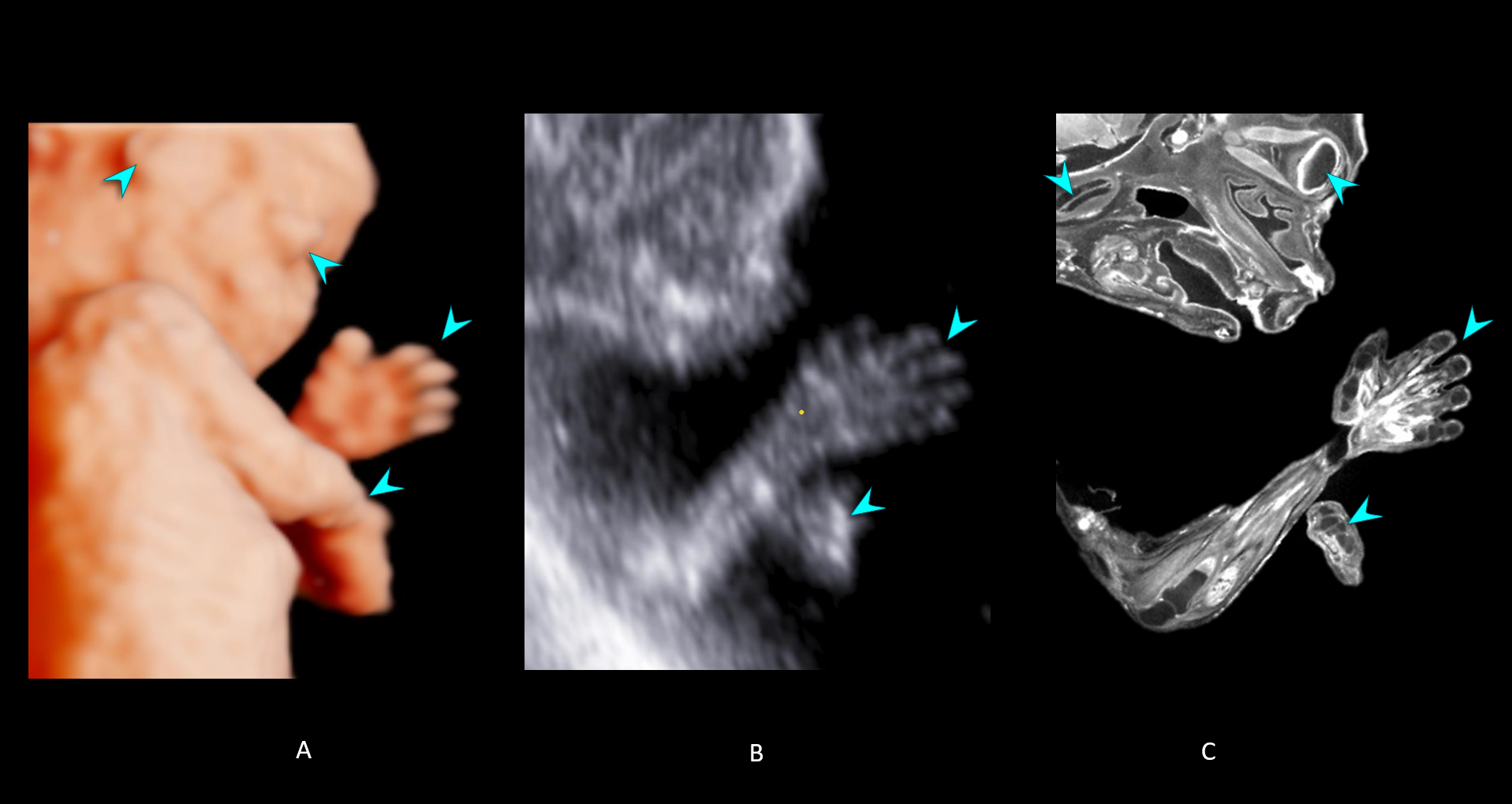
- 2D ultrasound image of the left hand from the 3D ultrasound volume, using the multiplanar mode.
- MicroCT image of a matching imaging plane selected from the same specimen.
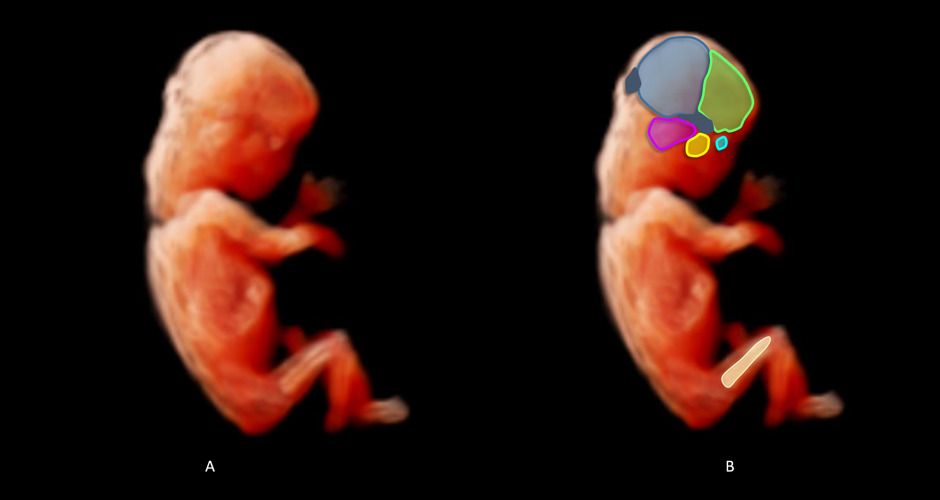
- 3D ultrasound volume with CrystalVue™ and RealisticVue™ applied.
- Overlay of a schematic illustration of the skeletal the structures visualised in the 3D ultrasound volume and the original image.
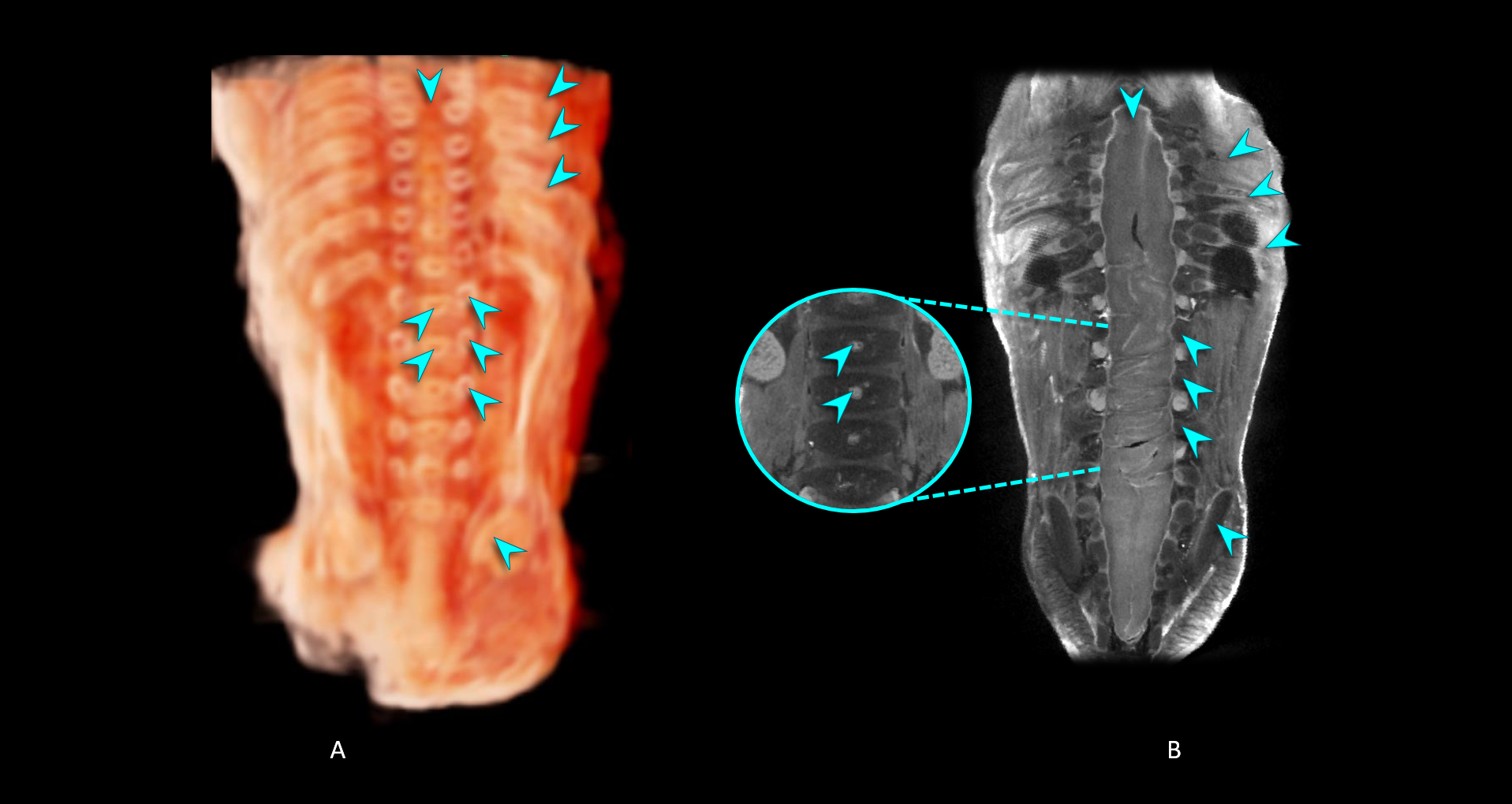
- 3D ultrasound volume with CrystalVue™ and RealisticVue™ applied. The volume has been sectioned in the coronal plane to visualise the fetal spine.
- MicroCT image slice corresponding to the 3D ultrasound section in image A to validate the the structures visualised*.
*Note that 3D imaging with CrystalVue™ rendering allows visualisation of multiple structures at different depths in a single imaging plane unlike microCT slices. As such, the vertebral bodies are visible in the ultrasound volume, whereas they are obstructed by the spinal cord in the microCT image even though the images have been acquired and displayed in the same imaging plane. Also note in image B that the spinal cord reaches all the way down to the bottom of the spinal canal at this stage of development.
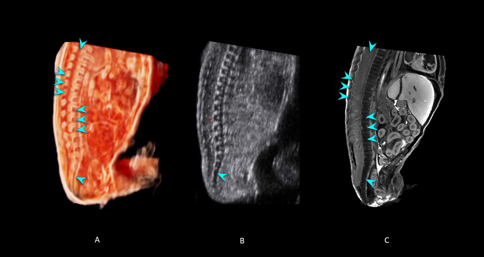
- 3D ultrasound volume with CrystalVue™ and RealisticVue™ rendering. The volume has been sectioned in the sagittal plane to visualise the fetal spine.
- 2D ultrasound image of the spine in the coronal plane from the 3D ultrasound volume, using the multiplanar mode.
- MicroCT image slice corresponding to the 3D ultrasound section in image A to validate the the structures visualised*.
*Note that the spinal cord appears translucent in the 3D ultrasound volume whereas the spinal cord can be clearly discerned in the microCT image. The spinal cord reaches down all the way down to the bottom of the spinal canal at this stage in development.
**Note that the sacral vertebrae do not fuse during fetal development.
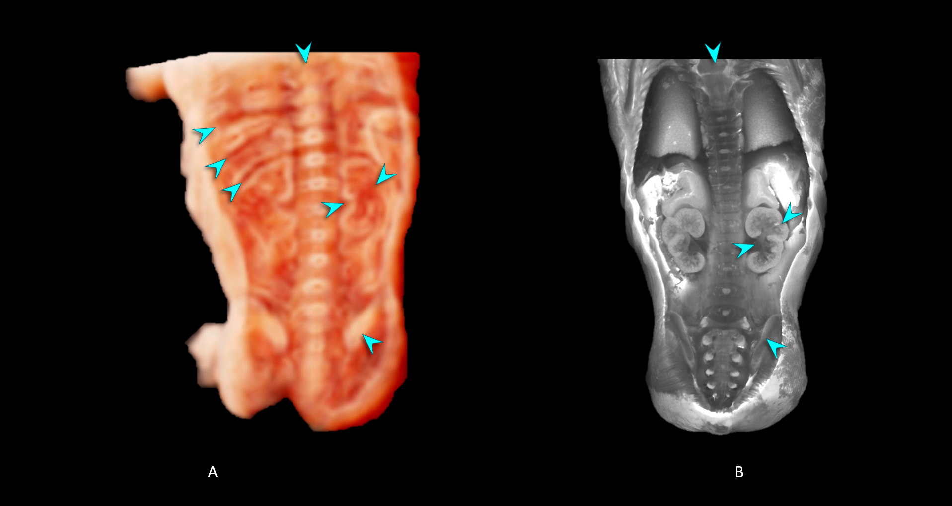
- 3D ultrasound volume with CrystalVue™ and RealisticVue™ rendering. The volume has been sectioned in a coronal plane to visualise kidneys.
- MicroCT image slice corresponding to the 3D ultrasound section in image.
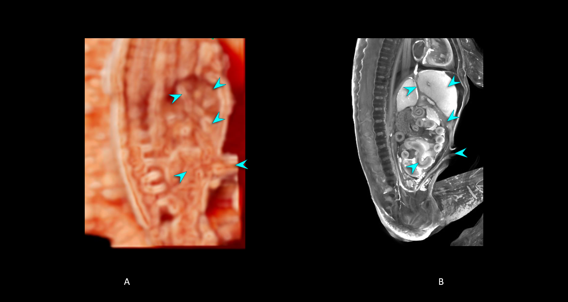
- 3D ultrasound volume with CrystalVue™ and RealisticVue™ rendering. The volume has been sectioned in a sagittal plane to visualise ductus venosus, umbilical vein and cord insertion.
- MicroCT image slice corresponding to the 3D ultrasound section in image A to validate the the structures visualised.
Comparative fetal anatomy
Case 2
Below are annotated 3D in-utero ultrasound images of a fetus – 13+1 weeks’ gestation – with CrystalVue™ and RealisticVue™ rendering as part of the PRECISE study. The ventricular system as shown on the 3D ultrasound volume was compared to a 3D model of the ventricular system of a Carnegie Stage (CS) 23 embryo, adapted from de Bakker et al., 2016, with permission.

- 3D ultrasound image in the coronal plane of the ventricular system at 13 weeks gestation with CrystalVue™ and RealisticVue™ rendering applied (in-utero volume from the PRECISE study).
- Digital reconstruction of the ventricular system of a stage 23 human embryo from the 3D Atlas of Human Embryology.
- 3D ultrasound image in the sagittal plane with CrystalVue™ and RealisticVue™ software applied.
- Schematic drawing of the ventricular system as presented in the sagittal plane.
H. Shah et al., Novel first-trimester fetal CNS ultrasonography. Am J Obstet Gynecol 2017.