Visualisation of extra-embryonic structures
Case 1
Below is an annotated image of a 14 weeks and 3 days (14 + 3 weeks) old fetus with Trisomy 21, born with the amniotic sac membrane intact. The specimen was imaged ex-vivo with 3D ultrasound with CrystalVue™ and RealisticVue™ rendering.
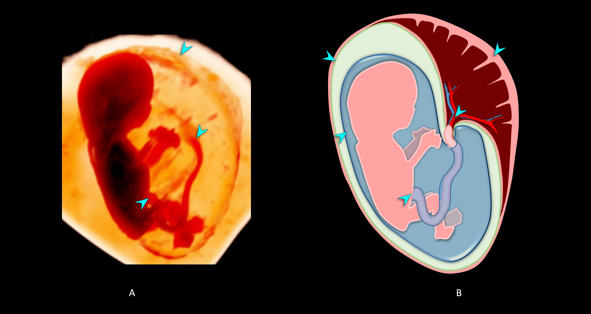
- 3D ultrasound volume with CrystalVue™ and RealisticVue™ applied of a fetus with Trisomy 21 at 14+3 weeks gestation, born with intact amniotic sac. The light source is placed behind the fetus enabling visualisation of the amniotic sac and insertions of the umbilical cord both into the placenta and anterior abdominal wall.
- Schematic illustration of the fetus and the fetal membranes. Structures with an asterisk [*] are not visible in the 3D ultrasound volume, but have been added to illustrate the relation between the developing fetus, fetal membranes, and placenta.
Comparative fetal anatomy
Case 2
Below are annotated images of a healthy fetus of 14+5 weeks gestation. This specimen was imaged ex-vivo using 3D ultrasound imaging with 3D ultrasound with CrystalVue™ and RealisticVue™ rendering. Thereafter, the specimen was imaged using contrast-enhanced microCT which was used as an anatomical reference standard. The ultrasound and microCT images are directly compared, allowing for validation and annotation of the the structures visualised in 3D ultrasound imaging.
The arrows and coloured fields in each image indicate anatomical structures. When you move your mouse over the arrow or coloured field, the name of the structure will appear.
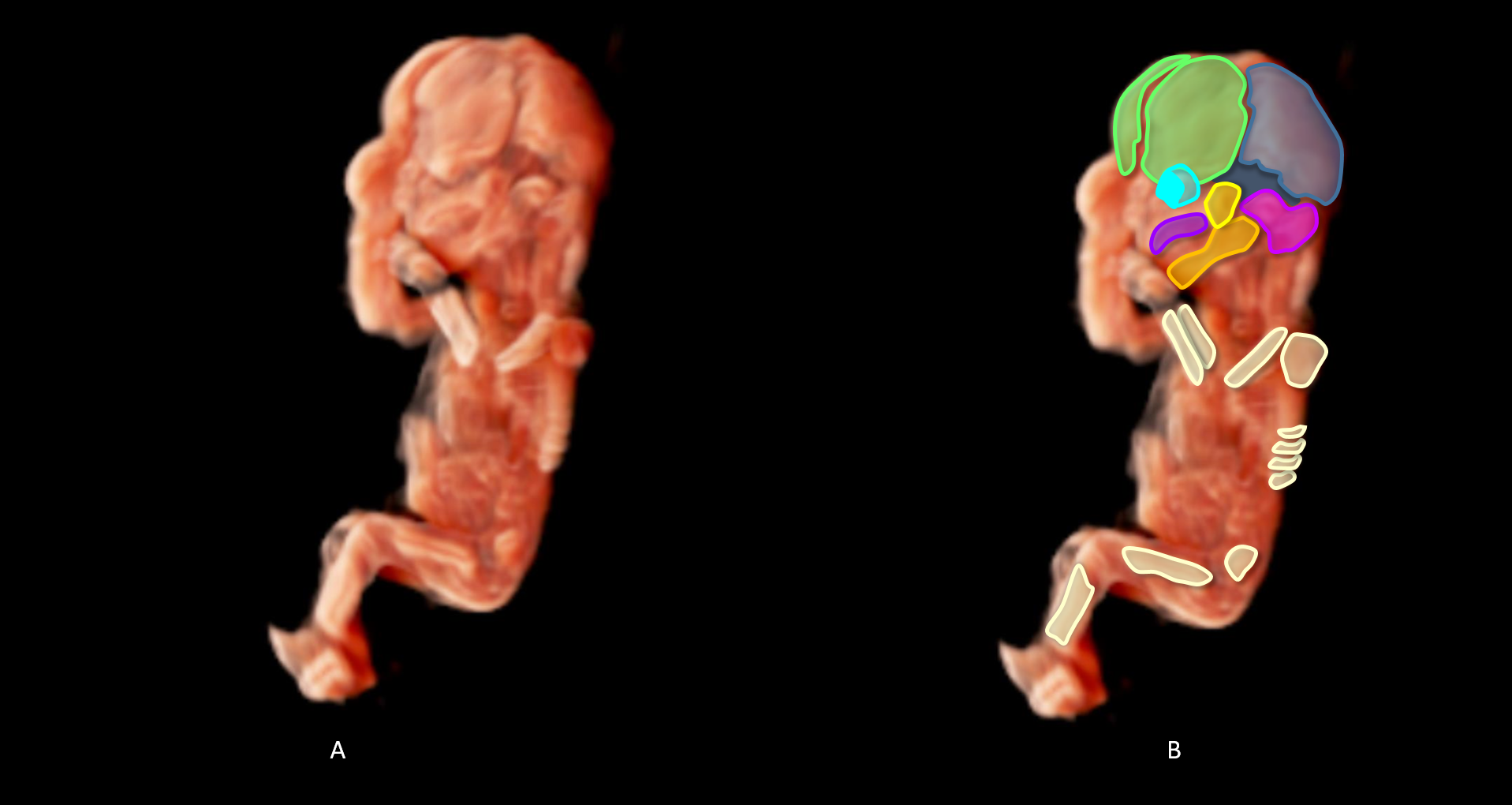
- 3D ultrasound volume with CrystalVue™ and RealisticVue™ applied. Settings are optimised for visualisation of skeletal structures.
- Overlay of a schematic illustration of the skeletal the structures visualised in the 3D ultrasound volume and the original image.
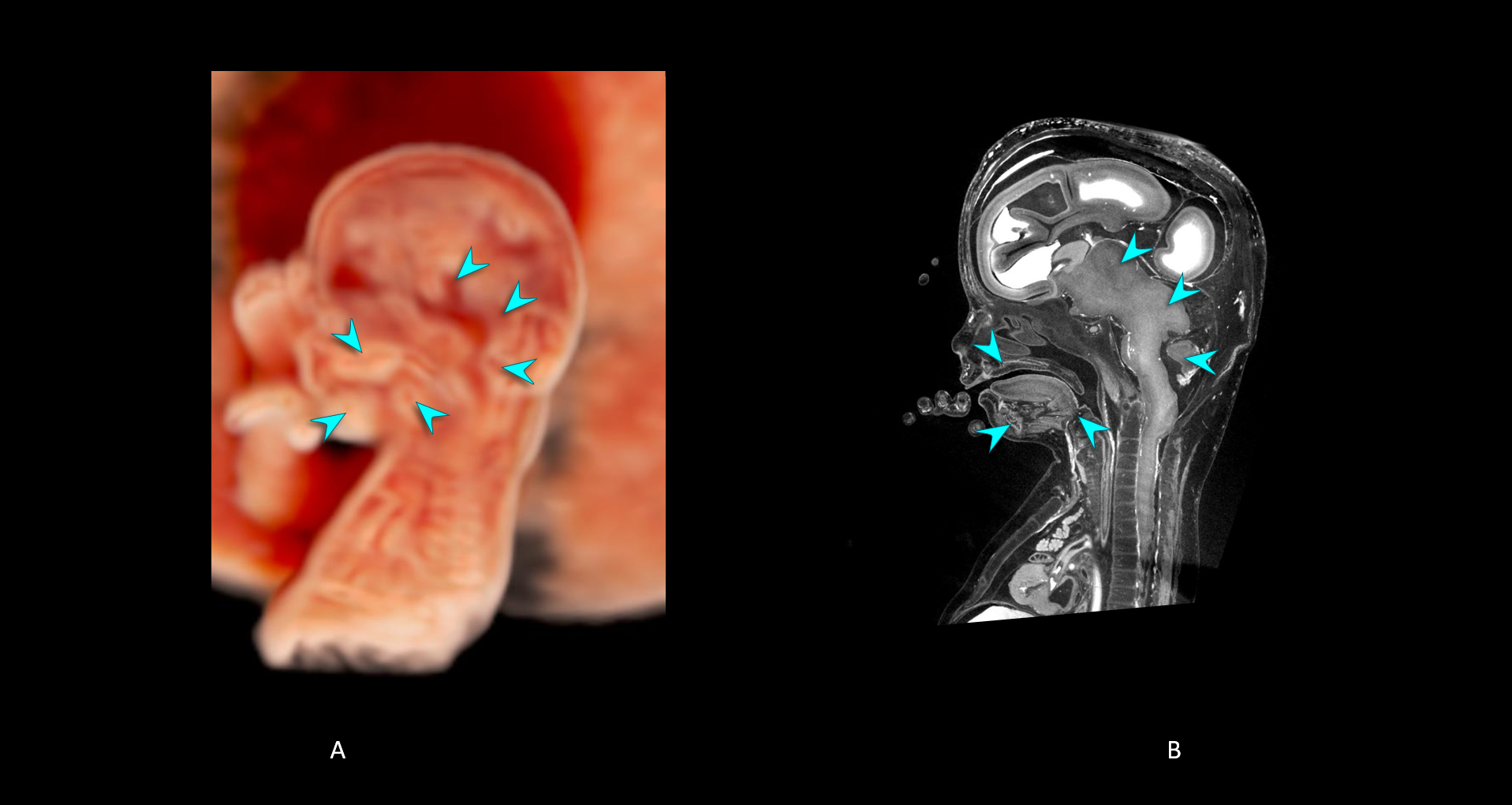
- 3D ultrasound volume with CrystalVue™ and RealisticVue™ applied. A mid sagittal view of the midline intracranial structures and palate is selected.
- MicroCT image slice corresponding to the 3D ultrasound section in image A to validate the the structures visualised.
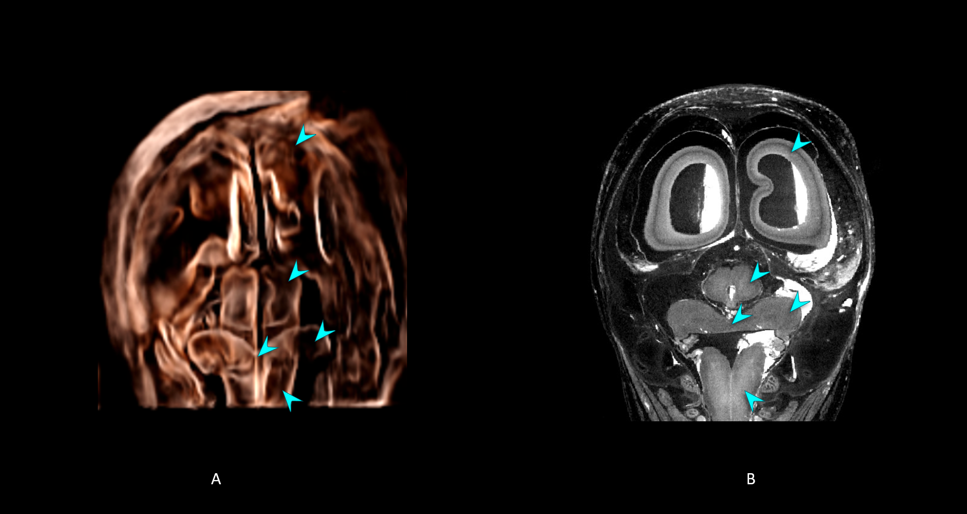
- 3D ultrasound volume with CrystalVue™ and RealisticVue™ applied. A coronal view of the posterior cranial fossa is selected.
- MicroCT image slice corresponding to the 3D ultrasound section in image A to validate the the structures visualised.
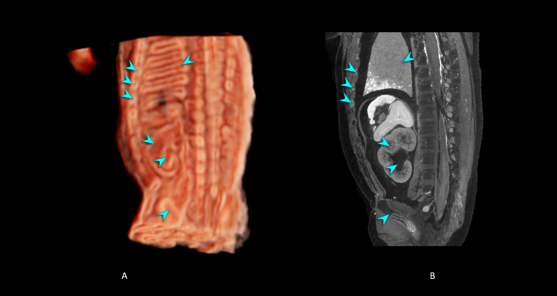
- 3D ultrasound volume with CrystalVue™ and RealisticVue™ applied. The volume has been sectioned through the coronal plane of the left kidney (note that the kidney is positioned slightly obliquely in the body with its pelvis facing towards the vertebral column).
- MicroCT image of the same specimen. A corresponding imaging plane was selected to validate the the structures visualised.
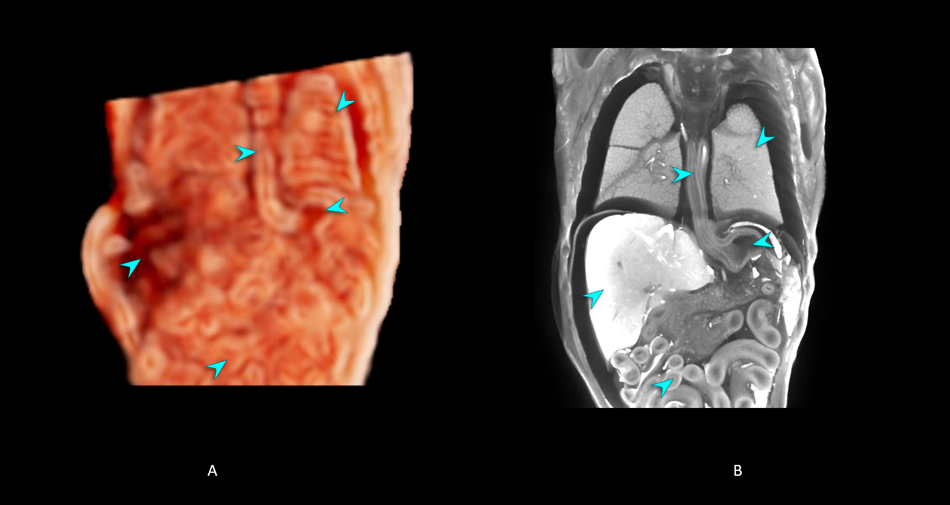
- 3D ultrasound volume with CrystalVue™ and RealisticVue™ applied. The volume has been sectioned through a coronal plane, visualising the distal oesophagus entering the stomach.
- MicroCT image of the same specimen. A corresponding imaging plane was selected to validate the the structures visualised.