Comparative fetal anatomy
Below are video clips of normal fetal anatomy at 12 weeks gestation, visualised by in-utero 3D ultrasound imaging with with CrystalVue™ and RealisticVue™ rendering applied. A healthy fetal specimen from the Carnegie Collection of a similar gestation and representing comparable development and morphology has been selected to compare the visualised anatomical structures.
How to use the video clips?
Start the video clips by pressing the play button. Click on the tabs to the left of the videos to view annotated still images from each clip.
3D ultrasound
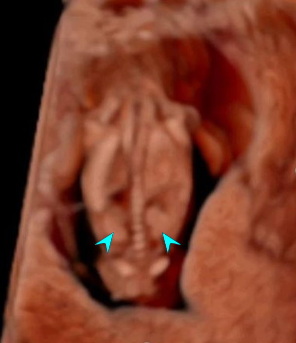
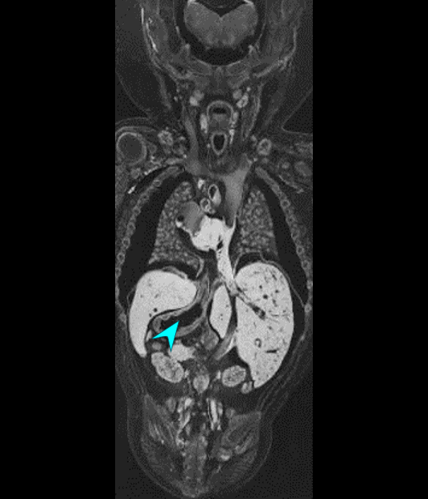
3D in-utero ultrasound volume of a healthy fetus at 12 weeks gestation, imaged as part of the PRECISE study. RealisticVue™ and CrystalVue™ rendering have been applied. The volume is sectioned in the coronal plane to visualise internal structures. Moving in a dorsal-ventral direction through the volume, structures such as the kidneys can be recognised. At 0:13, CrystalVue™ complexity is increased, making structures more see-through to enable visualisation of the stomach, lungs, liver and diaphragm. Adapted with permission from H. Shah, unpublished data.
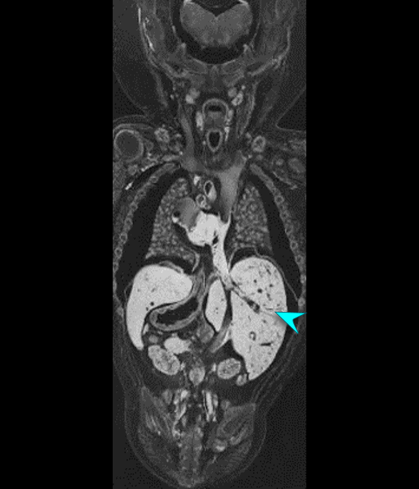
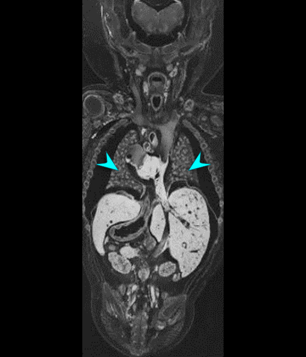
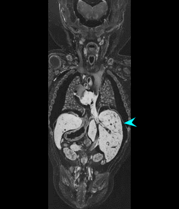
Histology
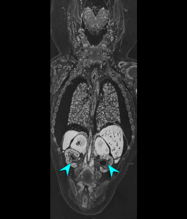
Histological sections of a fetal specimen from the Carnegie Collection, estimated at around 11 weeks gestation (CRL 42mm). The specimen is sectioned in the coronal plane, allowing for comparison of the anatomy visualised by 3D ultrasound imaging presented in the left pane. Adapted with permission from B.S. de Bakker, unpublished data.
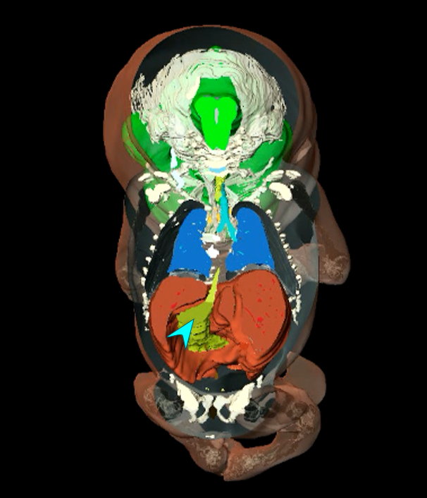
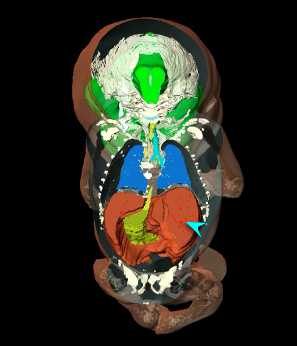
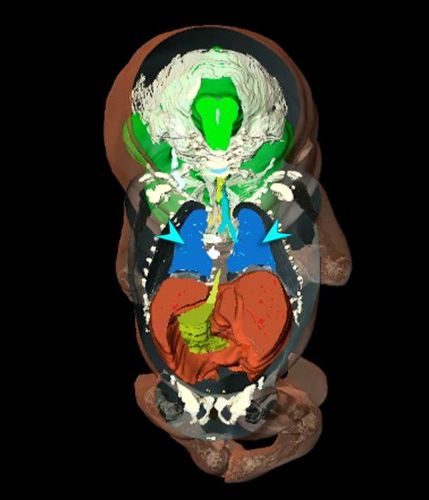
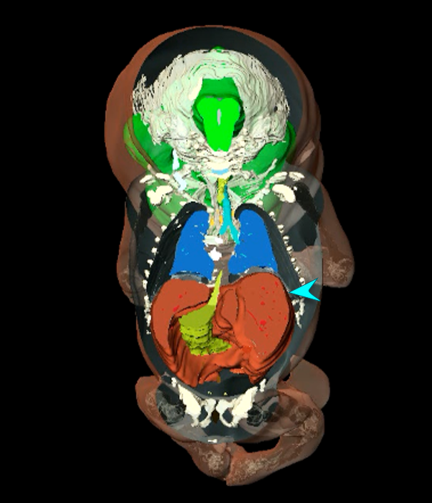
3D fetal anatomical model
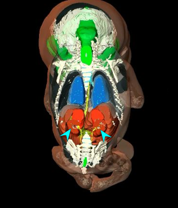
3D fetal model based on reconstruction of anatomical structures identified in the histological sections shown in the middle pane. Adapted with permission from B.S. de Bakker, unpublished data.