17 Weeks Gestation

3D Ultrasound Atlas

Comparative fetal anatomy
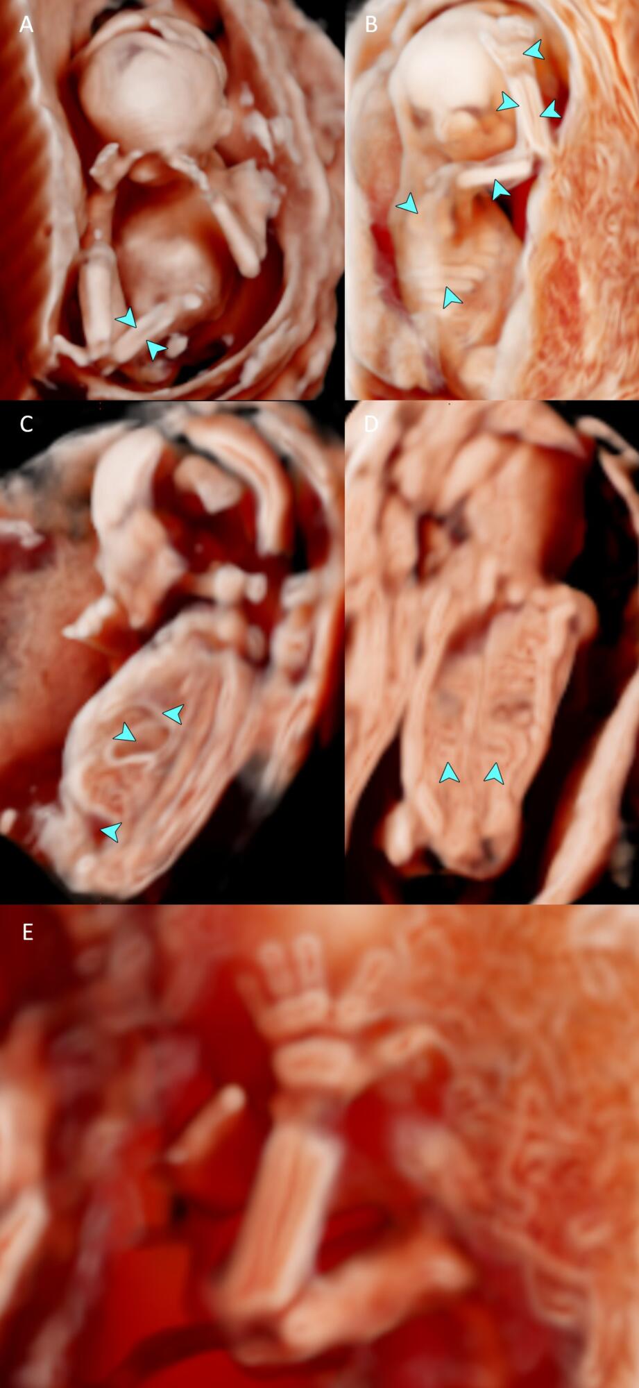
Below are annotated images of a healthy 16 weeks and 2 days (16 + 2 weeks) gestation fetus, imaged ex-vivo using 3D ultrasound with CrystalVue™ and RealisticVue™ rendering.

Thereafter, the specimen was imaged using contrast-enhanced microCT which was used as an anatomical reference standard. The ultrasound and microCT images are directly compared, allowing for validation and annotation of the the structures visualised in 3D ultrasound imaging. 

  1. – C. Increasing levels of CrystalVue™ complexity and transparency are applied to the same 3D ultrasound volume imaged with CrystalVue™ and RealisticVue™, highlighting different (internal) structures. See image below for detailed annotation of skeletal the structures visualised in image C.
  1. 3D ultrasound volume with CrystalVue™ and RealisticVue™ rendering. Settings are optimised to highlight the skeletal structures.
  2. Annotated skeletal structures.
  3. Overlay of a schematic illustration of the skeletal the structures visualised in the 3D ultrasound volume and the original image.
  1. 3D ultrasound volume CrystalVue™ and RealisticVue™ rendering of the head and brain. The volume has been inverted and sectioned in a parasagittal plane to visualise the midbrain and right hemisphere. For this volume, a high-frequency transducer was used (3-14 MHz).
  2. MicroCT image of the same specimen. A corresponding imaging plane was selected to validate the the structures visualised.
  3.  3D ultrasound volume of the complete specimen, imaged using low frequency transducer (1-8MHz). The volume has been inverted and the level of CrystalVue™ complexity and transparency have been increased, creating a silhouette of the fetus and intracranial structures.
  1. 3D ultrasound volume CrystalVue™ and RealisticVue™ rendering, imaged using a low frequency (1-8 MHz) transducer. The volume has been inverted and sectioned in a parasagittal plane. Midline brain structures and the anterior horn of the lateral ventricle are visualised in this view.
  2. MicroCT image of the same specimen. A corresponding para-sagittal imaging plane has been selected to validate the structures visualised.
  1. 3D ultrasound volume with CrystalVue™ and RealisticVue™ applied. A mid sagittal view of the head is selected, visualising the palate as well as some midline brain structures.
  2. MicroCT image slice corresponding to the 3D ultrasound section in image A to validate the the structures visualised.
  1. 3D ultrasound volume with CrystalVue™ and RealisticVue™ applied. A coronal plane of the posterior cranial fossa displays the cerebral hemispheres, thalamus, midbrain, cerebellar peduncles and brainstem.
  2. MicroCT image slice corresponding to the 3D ultrasound section in image A to validate the the structures visualised.
  1. 3D ultrasound volume with CrystalVue™ and RealisticVue™ rendering. The volume has been sectioned in the coronal plane, allowing three-dimensional visualisation of the lungs, diaphragm, lever and stomach.
  2. MicroCT image of the same specimen. A matching coronal plane was selected to validate the structures visualised.
  3. Overlay of a schematic illustration of chest and abdominal organs visualised in the 3D ultrasound volume and the original figure.