3D Ultrasound Atlas
Comparative fetal anatomy
Below are annotated images of a fetus at 10 weeks gestation, imaged in-vivo using 3D ultrasound imaging with CrystalVue™ and RealisticVue™ applied.
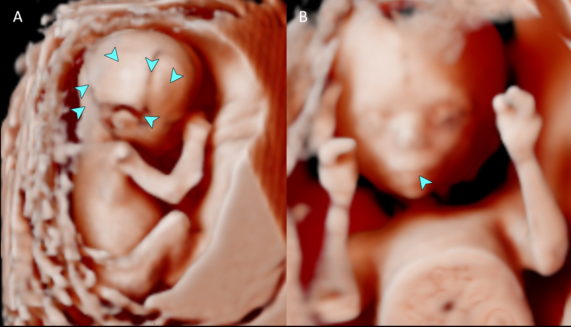
Skull bones, sutures and features of the fetal face including the lips at 15 weeks gestation.
3D ultrasound volume with CrystalVue™ and RealisticVue™ rendering.
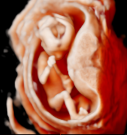
Fetal limbs including hands and feet.
You can see into the brain here with CrystalVue – falx ands ventricle
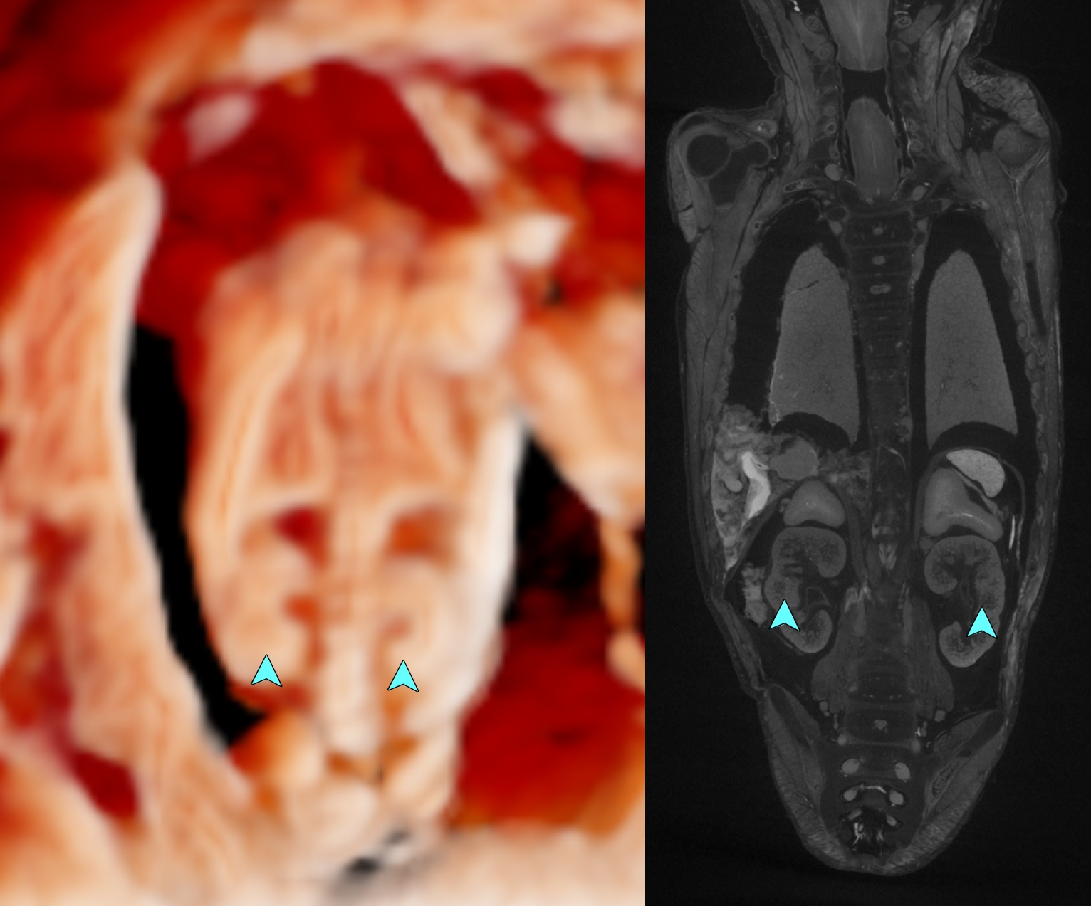
3D ultrasound volume with CrystalVue™ and RealisticVue™ rendering, visualising the kidneys in-vivo. The 3D ultrasound image is compared to specimen TOP 62 (15+0) from the Dutch Fetal Biobank.
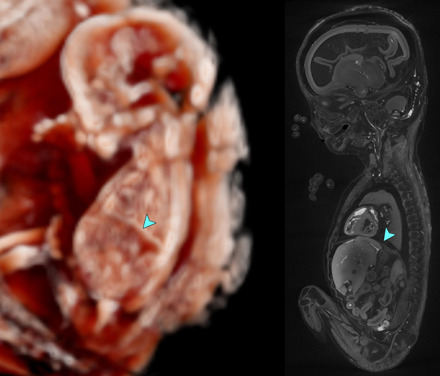
3D ultrasound volume with CrystalVue™ and RealisticVue™ rendering, visualising the diaphragm in-vivo. The 3D ultrasound image is compared to specimen TOP 62 (15+0) from the Dutch Fetal Biobank.